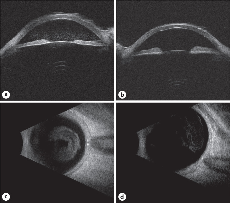
Fig. 8.
Ultrasound imaging for case 3. Ultrasound biomicroscopy demonstrated anterior and posterior chamber debris in the right eye (a), while only posterior chamber debris in the left eye (b). B-scan imaging of the right (c) and left eye (d) showed dense vitreous opacities, right greater than left.

