Fig. 9.

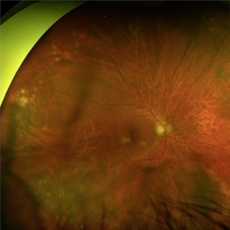
Fundus image of case 4. Color fundus photo of the right eye with dense syneresis. Posteriorly, there was a Weiss ring and lattice degeneration without tears or detachments. A large choroidal nevus was seen in the inferonasal periphery.

Fundus image of case 4. Color fundus photo of the right eye with dense syneresis. Posteriorly, there was a Weiss ring and lattice degeneration without tears or detachments. A large choroidal nevus was seen in the inferonasal periphery.