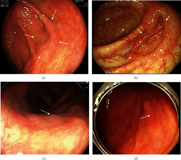
Figure 2.

Endoscopic finding of SSL and SSLD with WLI. (a) WLI finding of SSL. A fading and slightly elevated lesion of 20 mm (white arrows). (b) WLI finding of SSLD. A lesion of 15 mm with central depression (white arrows). (c) WLI finding of SSLD. A lesion of 18 mm with a granular elevation (white arrow). (d) WLI finding of SSLD. A lesion of 18 mm with partial redness (white arrow).
