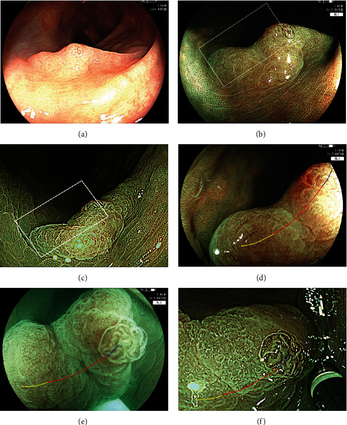
Figure 4.

A case presentation of SSLD with WDC. (a) A SSLD of 15 mm on the descending colon with WLI. It is a fading and slightly depressed lesion with a granular elevation. (b) Image with BLI. (c) Image with NBI. (d) BLI magnification of the white box in (b). NV is observed on the granule (blue line). WDC is observed on the depressed area (red line). WDC is not observed (yellow line). (e) BLI magnification of white box under water. WDC appears whiter and clearer than the regular view under air. (f) NBI magnification of the white box in (c). NV is observed on the granule (blue line). White dendritic change is observed on the depressed area (red line), and WDC is not observed (yellow line).
