
Figure 5.
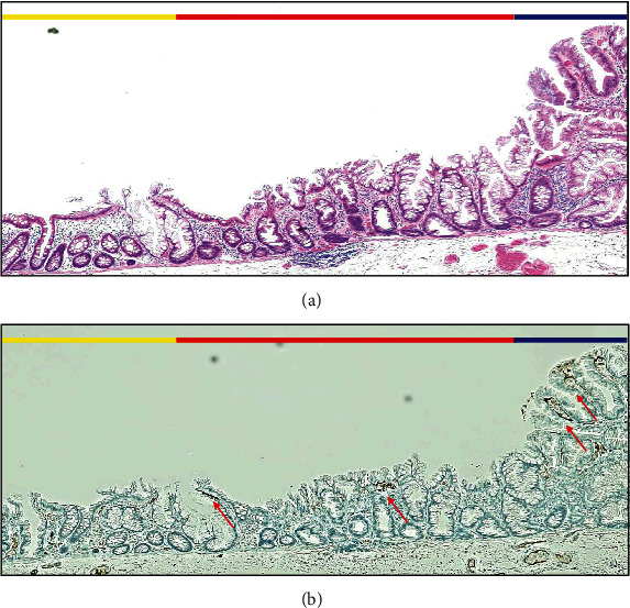
Histopathological findings of WDC in H.E. stains and immunohistochemical examination. (a) Histopathological examination with H.E. stains of blue, red, and yellow lines in Figure 4(c). The blue line corresponds to the area of dysplasia, and it does not show WDC. The red line corresponds to WDC. The yellow line corresponds to an area without WDC. In the area of WDC (red line), serrated-shaped superficial epithelium of the colonic mucosa is observed. (b) Immunohistochemical examination with adipophilin of (a) shows several minor positive areas (red arrows). In WDC area (red line), positive of adipophilin is detected. But it is also detected in the area of dysplasia (blue line).