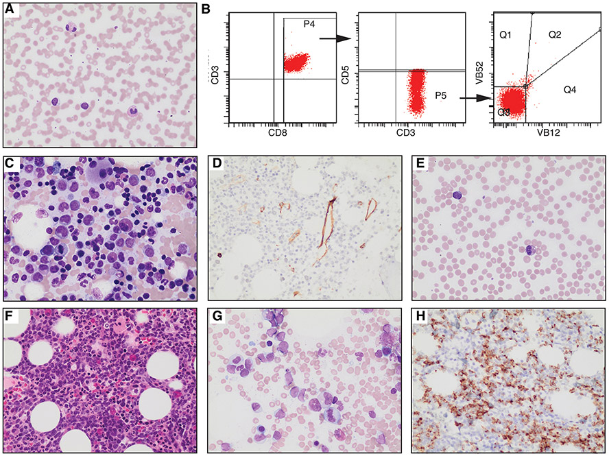
Figure 1. Morphological and immunophenotypic characterization of T-LGLL and Subsequent AML.
A-D, samples from 2018 specimens. E-H, samples from 2022 specimens. (A) Peripheral blood smear of T-LGLL (Giemsa stain, 500X). Increased eosinophils and small, atypical lymphocytes with cytoplasmic granules are observed. (B) Flow cytometry of the patient’s peripheral blood showed atypical T cells [CD3+, CD8+, CD5dim/−, CD7dim/− (not shown), and Vbeta-]. (C) Bone marrow aspirate (C: 500X) shows scattered small lymphocytes, increased eosinophils, and dysplastic megakaryocytes. (D) Immunohistochemical staining for CD34 (400X). (E) Peripheral blood smear (Giemsa stain, 500X) showing increased eosinophils and occasional circulating myeloblasts. (F) Bone marrow core biopsy (400X) exhibiting hypercellularity with a left shift. (G) Bone marrow aspirate (500X) indicating elevated myeloblasts. (H) CD34 immunostaining of the bone marrow core (400X).

