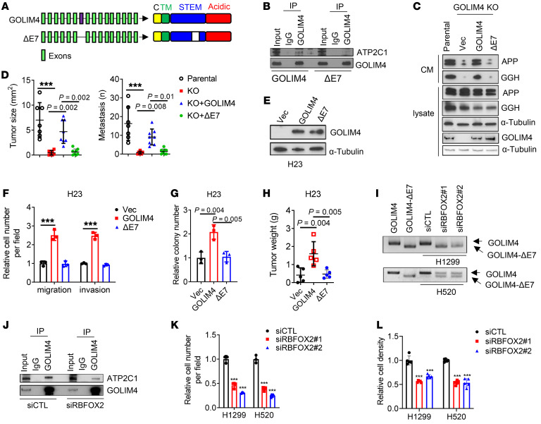
Figure 5. An alternatively spliced exon in GOLIM4 is required for ATP2C1-binding and secretory activities.
(A) Full-length and spliced (ΔE7) GOLIM4 isoforms. Exon 7 is located in the intraluminal STEM domain. TM, transmembrane domain; C, cytoplasmic domain. (B) IP/WB analysis of GOLIM4-KO H1299 cells reconstituted with full-length or ΔE7 GOLIM4. ATP2C1-binding activity was detected only in full-length GOLIM4-transfected cells. (C) WB analysis of secreted proteins in CM samples and cell lysates from parental and GOLIM4-KO H1299 cells reconstituted with full-length or ΔE7 GOLIM4. α-Tubulin was used as a loading control. (D) Quantification of orthotopic tumor size (left) and distant metastases (right) per mouse (data points) generated by H1299 cells in C. (E) WB analysis of GOLIM4 levels in H23 cells transfected with full-length GOLIM4 or GOLIM4-ΔE7. (F–H) Boyden chamber migration and invasion assays (F), soft agar colony assays (G), and flank tumor growth assays (H) on cells in E. (I) qRT-PCR analysis of GOLIM4 isoforms in siRNA-transfected H1299 and H520 cells. Full-length GOLIM4 and GOLIM4-ΔE7 were included as controls. (J) IP/WB analysis of H1299 cells demonstrates that FOXF2 depletion attenuated the ATP2C1-binding activity of GOLIM4. (K and L) Boyden chamber migration assays (K) and relative cell density assays (L) on siRNA-transfected H1299 and H520 cells. Data indicate the mean ± SD from a single experiment incorporating biological replicate samples (n = 3, unless otherwise indicated) and are representative of at least 2 independent experiments. ***P < 0.001, by 1-way ANOVA (D, F–H, K, and L).

