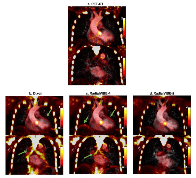
Fig. 2.
Thoracic sodium [18F]fluoride uptake in two coronal planes in a patient with bicuspid aortic valve. (a) Positron emission tomography (PET)-computed tomography (CT) co-registered with magnetic resonance angiography (MRA), (b) combined Dixon attenuation correction PET and MRA, (c) combined RadialVIBE-4 attenuation correction PET and MRA, and (d) combined RadialVIBE-2 attenuation correction PET and MRA. Sodium [18F]fluoride uptake in each PET-MRI image demonstrates a similar pattern compared with uptake in PET-CT. Note the image artefact in Dixon and RadialVIBE-4 at the bronchial tree and heart-lung and diaphragm-lung border (green arrows). Colour scale on the right of images represents standardised uptake values

