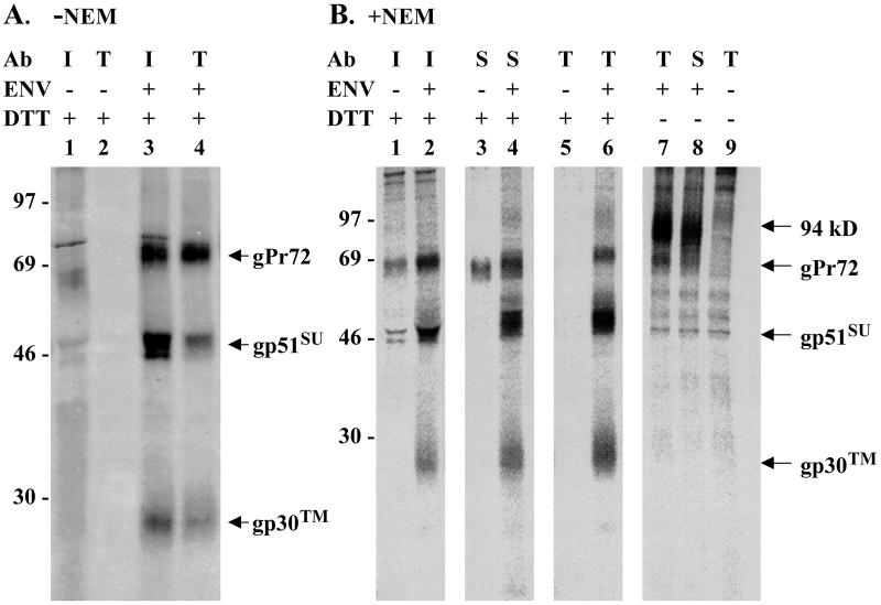
FIG. 2.
Disulfide linkage of SU and TM proteins in transfected cells overexpressing BLV envelope protein. COS-1 cells transfected with a BLV Env expression vector (+) or a CAT expression vector (−) were radiolabeled with [35S]cysteine, and glycoproteins concentrated from cell lysates were subjected to immunoprecipitation. (A) Cells were lysed in the absence of NEM. Glycoproteins recovered from 2 × 10⁵ transfected cells were precipitated with serum (diluted 1:266) from a BLV-infected sheep (I), and the supernatants were then precipitated with rabbit anti-TM (T) antibody (Ab) diluted 1:1,000. Bound proteins were eluted from protein G- or A-Sepharose with double-strength SDS gel sample buffer containing 100 mM DTT. The eluates were boiled for 4 min, and proteins were separated by electrophoresis on SDS–12% polyacrylamide gels. The gel was fixed with 30% methanol–10% acetic acid, impregnated with En3Hance (DuPont), dried, and then exposed to X-ray film for 14 days. The developed film was scanned at 300 pixels/in., and the image was printed using Adobe Photoshop. (B) Before being lysed in buffer containing 10 mM NEM, cells were incubated in cold phosphate-buffered saline containing 10 mM NEM. Each lane represents glycoproteins from 105 transfected cells that were present in immunoprecipitates obtained using infected sheep serum (I) diluted 1:133, monoclonal antibody specific for the D epitope of SU (S) diluted 1:200, or anti-TM antibody (T) diluted 1:1,000. Prior to electrophoresis, precipitated proteins were heated with or without DTT, as indicated. A PhosphorImager screen was exposed to the fluor-impregnated gel for 4 days before being scanned on a Storm 860 (Molecular Dynamics). Images digitized using Image-Quant software were printed using Adobe Photoshop. The values to the left are molecular sizes in kilodaltons.

