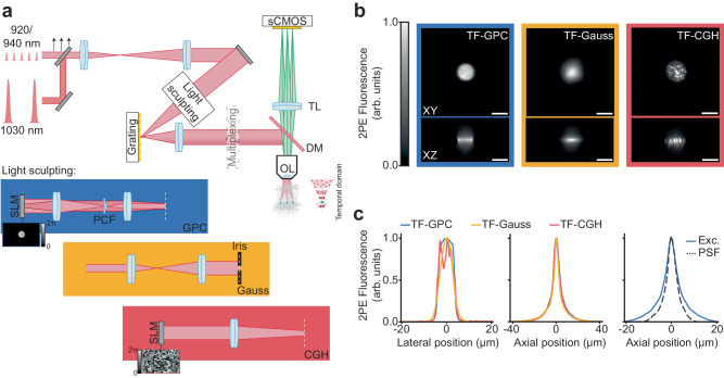
Fig. 1. Characterisation of the optical setup developed for scanless two-photon voltage imaging.
a Schematic diagram of a typical optical setup used to perform scanless 2P voltage imaging (refer also to Supplementary Fig. 1). High repetition rate (920 or 940 nm) sources delivering nJ pulses at 80 MHz and fixed wavelength (1030 nm) low repetition rate sources delivering µJ pulses (at variable repetition rates) were used for scanless 2P voltage imaging. The black arrows indicate an adjustable mirror used to direct light from either ultrafast source into the microscope. The light sculpting paths were designed to generate Temporally Focused (TF), Generalised Phase Contrast (GPC), Gaussian and holographic (CGH) spots with lateral FWHM (Full Width Half Maximum) diameters between 12 and 17 µm. The essential concepts of each light sculpting approach are depicted in the inset below the schematic. Diffraction gratings were positioned in conjugate image planes for temporal focusing, as indicated by the white dashed lines. Temporal focusing was used to maintain axial resolution in spite of the extended lateral spot size. 2P excited fluorescence was collected using a widefield detection axis equipped with an sCMOS camera capable of kilohertz acquisition rates. Patch-clamp electrophysiology apparatus was installed on each microscope (“Methods” section). OL objective lens, DM dichroic mirror, TL tube lens, sCMOS scientific complementary metal–oxide–semiconductor, SLM spatial light modulator, PCF phase contrast filter. b Lateral (XY) and axial (Z) cross-sections of representative 2P excited fluorescence generated in a thin spin-coated rhodamine layer with 12 µm TF-GPC, TF-Gaussian and TF-CGH spots, as indicated. Scale bars represent 10 µm. c Lateral and axial profiles of 2P excited fluorescence generated with each excitation modality, and the corresponding system response. Exc. refers to the average excitation axial profile for all modalities and PSF to the effective Point Spread Function of scanless 2P microscopy, as measured using 1 µm fluorescence microspheres excited at 940 nm with TF-GPC. Source data are provided as a Source Data file.

