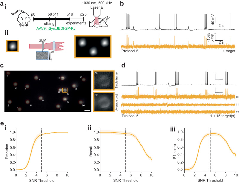
Fig. 5. Multi-target scanless two-photon voltage imaging using low repetition rate sources at 1030 nm.
a (i) Schematic representation of the protocol used to prepare JEDI-2P-Kv expressing hippocampal organotypic slices (see “Methods” section, Supplementary Table 2). (ii) Multiple cells were illuminated simultaneously by multiplexing a temporally focused (TF) Gaussian beam with a Spatial Light Modulator (SLM, “Methods”, Supplementary Table 2). All experiments were performed using 17 µm TF-Gaussian spots at 1030 nm (laser E, 500 kHz repetition rate, power densities: 0.03–0.06 mW µm−2). Data was acquired for 30 s at an acquisition rate of 500 Hz with camera A. b Simultaneous current-clamp (upper, black) and fluorescence recordings (lower, yellow) of electrically evoked activity in neurons from hippocampal organotypic slices (protocol 5, “Methods” and Supplementary Table 5). c Average (temporal) projection of data acquired during a representative multi-target scanless 2P voltage imaging experiment (grey lookup-table). The spot positions have been overlayed (yellow lookup-table) and numbered 1-16. The scale bar represents 20 µm. The patched neuron (cell 10) is indicated by the yellow box. Inset: single frame and average temporal projection of a zoomed portion of the dataset containing the patched cell. d Simultaneous current-clamp (upper, black) and fluorescence recordings (lower, yellow) of electrically evoked activity in neurons from hippocampal organotypic slices. Data acquired by illuminating the same patched cell plus fifteen randomly distributed positions (1 + 15) as shown in c. The number to the left of each trace indicates the index of the targeted cell as labelled in c. e (i) Precision, (ii) recall and (iii) F1 score of action potential detection plotted as a function of SNR threshold. The 95% confidence intervals are also plotted (shaded region). Refer to the “Methods” section for definitions of these terms. Simultaneously acquired whole-cell patch-clamp electrophysiology traces were used as ground-truth datasets. Of 380 electrically evoked action potentials, 96% were detected. The SNR threshold of 5, used throughout this manuscript, is indicated by the black dashed line (n = 15 neurons, from 3 different slices from 2 independent slice cultures). Source data are provided as a Source Data file.

