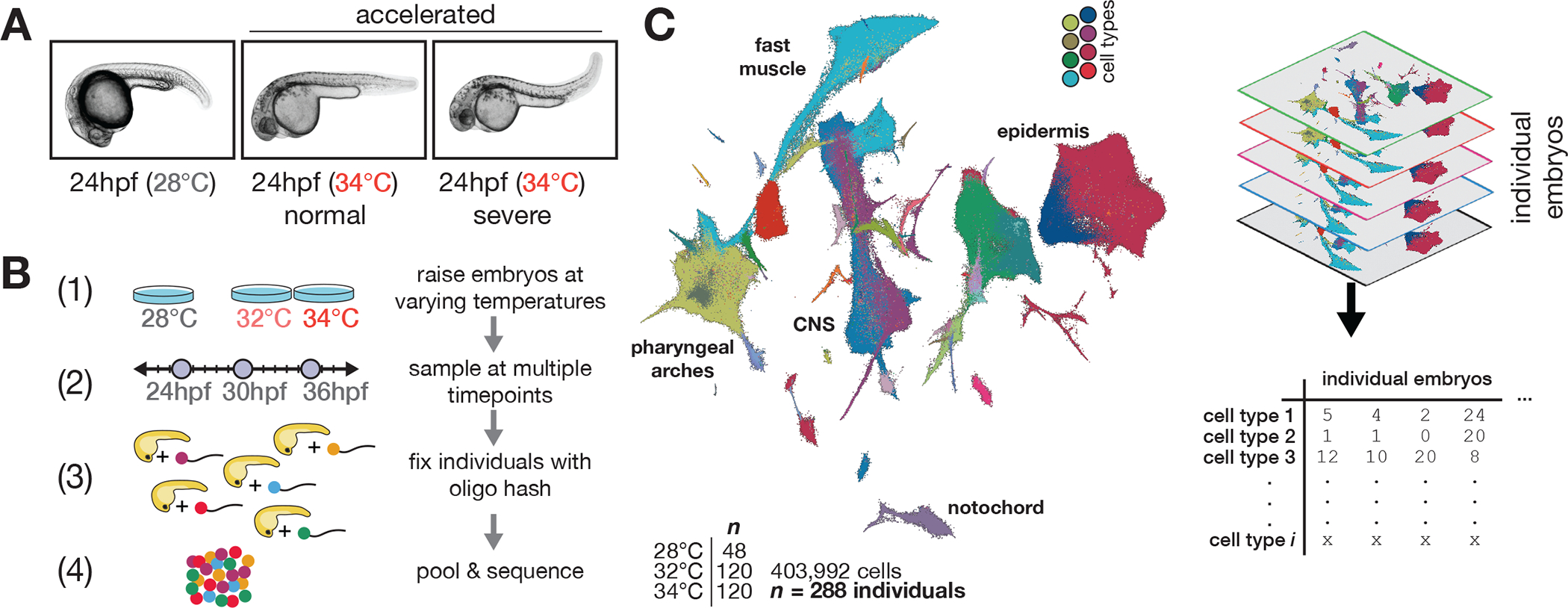
Figure 1. Effects of stress on phenotypic variability is captured via individual animal hashing of single-cell transcriptomes.

(A), Representative images of 24 hpf embryos raised at standard and elevated temperature; individual embryos with normal-looking and bent-tail phenotypes were included in the dataset. (B), Experimental workflow for temperature perturbation experiment and individual embryo hashing. (C), UMAP of temperature-perturbation dataset, projected into coordinate space of reference atlas (see related manuscript, 32). Right side shows how single cell data are transformed to generate cell composition matrices.
