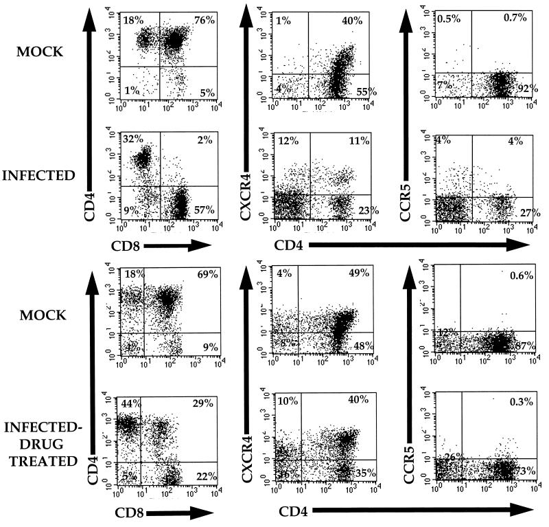
FIG. 1.
Effects of HAART on thymocytes. SCID-hu mice were constructed, as described previously, at the University of California–Los Angeles (1, 12). Thy/Liv implants were infected with HIV-1NL4-3, and biopsies were taken at 8 weeks (A) and 11 weeks (B) postinfection and analyzed by flow cytometry. Immediately following the biopsies 8 weeks postinfection, mice were administered a daily combination of AZT, ddI and indinavir for the remainder of the experiment. Cells at each time point were stained with antibodies specific for CCR5 (FITC), CXCR4 (PE), CD4 (Red613), and CD8 (APC). The panels show flow cytometry profiles of a representative mock-infected animal (animal no. 175-28, top row) and the flow cytometry profiles of an HIV-1NL4-3-infected animal (animal no. 175-29, lower row). Percentages of cells within each relevant quadrant are provided.

