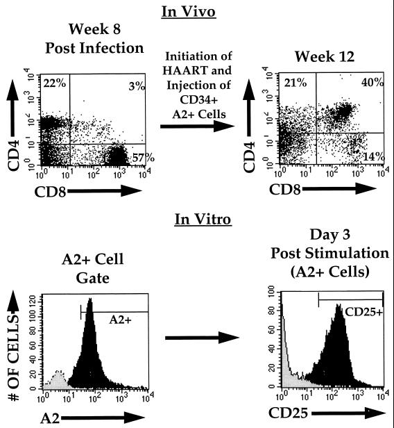
FIG. 3.
Functional response of the reconstituting thymocyte population. HIV-infected or mock-infected (not shown) thymic implants were biopsied 8 weeks postinfection and analyzed by flow cytometry for CD4 (biotin-Red613) and CD8 (APC) (upper left panel). HAART was initiated at this time, and CD34+/HLA-A2+ cells were injected into the implants at 8.5 weeks postinfection. A second biopsy was performed at 12 weeks postinfection, at which time cells were examined by flow cytometry (upper right panel) and cultured in the presence or absence of costimulation. Three days following costimulation, cells derived from implants not receiving HLA-A2+ cells (grey histogram) and those receiving HLA-A2+ CD34+ cells (black histogram) were analyzed for HLA-A2 (FITC) expression (lower left panel). HLA-A2+ cells in the implant, which in this mouse (animal no. 179-28) constituted the majority of cells, were gated and analyzed for CD25 (interleukin-2 receptor) (PE) expression (lower right panel) (black histogram). Unstimulated cells were cultured and analyzed in parallel (grey histogram). At this time, 92% of costimulated cells and 1% of unstimulated cells expressed CD25. CD25 expression was less than 1% in freshly isolated thymocytes prior to stimulation (not shown). Similar expression of CD25 was seen following costimulation of thymocytes from two mock-infected and six HIV-infected mice.

