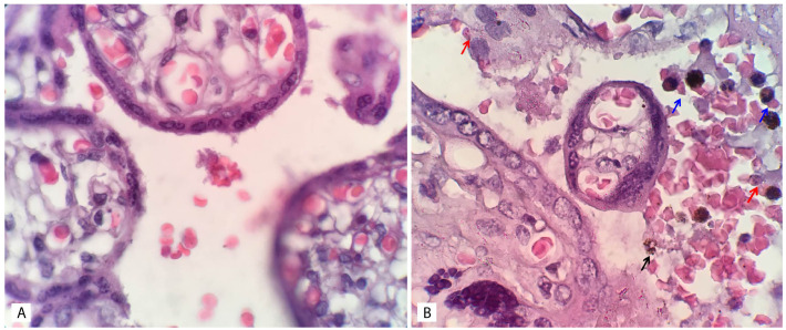
Figure 2. Histopathological examination of placental tissue.
A: Normal placental tissue. B: Placental malaria. The red arrow indicates infected erythrocyte sequestration, the blue arrow indicates monocyte infiltration, and the black arrow indicates free hemozoin in the intervillous space. The image size is the only modification made; no changes to brightness or contrast have been made. The examination was performed by using a light microscope (Olympus Biological Microscope, CX23) with 1000x magnification. The figures were captured using a mobile device, which could explain the absence of a scale bar.

