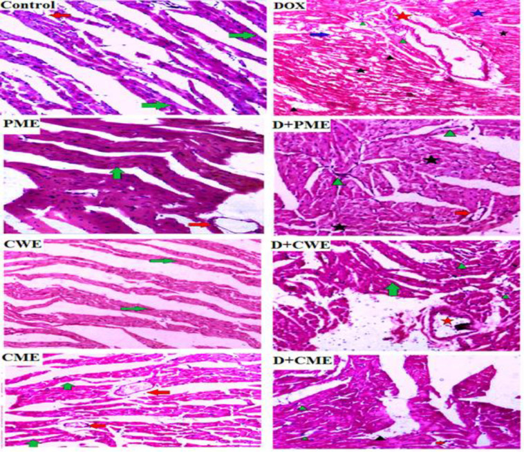
Figure 6.
Influence of different treatments on the heart tissue (H&E staining, 20X magnification). The myocardial fibers in the control image are normal in appearance and arrangement (green arrow), and a typical blood vessel (red arrow) is shown with them. DOX, on the other hand, shows vacuolated myocardial fibers with the disordered arrangement (blue arrow), an obviously thickened blood vessel with an elongated wall (red star), hyperemia (black arrow-head), infiltration of fibroblasts and lymphatic cells (green arrow-head); D+PME, it identify a regular blood vessel (red arrow), a little amount of fibroblast infiltration (green arrowhead), and minor myocytolysis (black star); PME demonstrates the typical appearance of the myocardial fibers (green arrow) with regular blood vessels (red arrow); Normal cardiac fibers are revealed by D+CWE (green arrow), along with a very little amount of inflammatory cells (green arrowhead), and a mildly dilated blood vessel with a thicker wall (red star). CWE shows a blood vessel with a typical appearance (red arrow), D+CME shows a blood vessel with a typical appearance (red arrow), mild lymphatic and fibroblast infiltration (green arrowhead), and hyperemia (black arrowhead), and CME shows the normal myocardial fibers appearance (green arrow) with healthy blood vessel (red arrow)

