FIGURE 1.
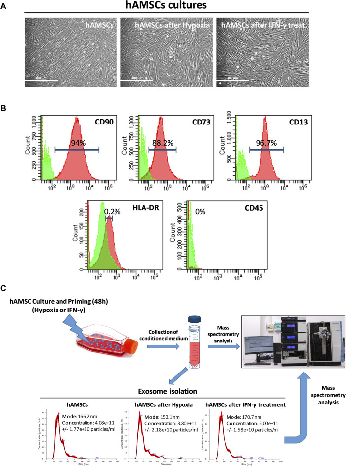
Human amnion-derived mesenchymal stromal/stem cells (hAMSCs) were grown as monolayers with or without priming. (A) Representative DIC images of hAMSCs grown as monolayers without priming (hAMSCs), cultured under hypoxic conditions (hAMSCs after hypoxia), or treated with IFN-γ (hAMSCs after IFN-γ treatment). (B) Representative images of flow cytometry analysis for quantification of hAMSCs at step 2 for both positive (CD90, CD73 and CD13) and negative surface markers (HLA-DR and CD45). Green represents the isotype control, and red represents stained cells. (C) Experimental plan and exosome characterization (size and concentration). DIC, differential interference contrast.

