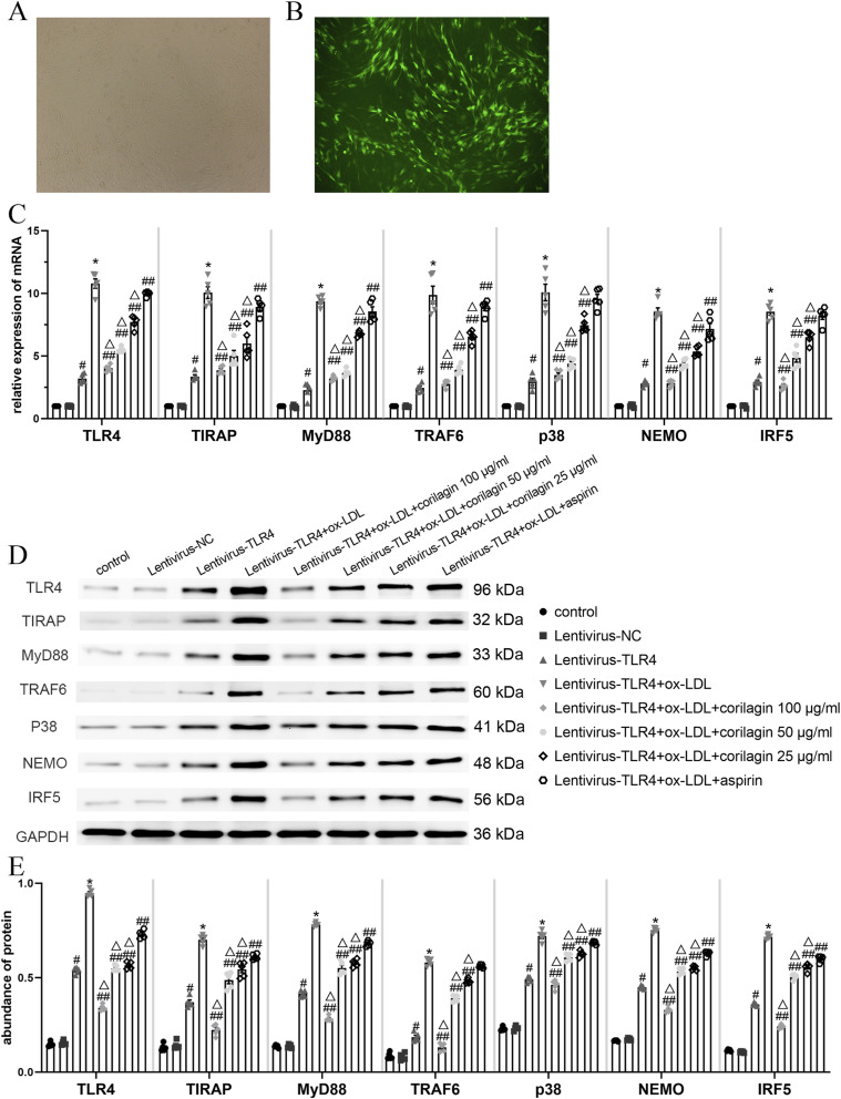
Figure 4.
Effect of corilagin on the TLR4 signaling pathway after upregulation of TL4 expression in MOVAS cells stimulated by ox-LDL. (A. B) 72 hours after transfection of the lentiviral vector, >85% of MOVAS cells expressed green fluorescent protein as observed with a fluorescence microscope. (C) mRNA expression was measured by qRT-PCR. #p < .05 compared with the control group, *p < .05 compared with the lentivirus-TLR4 group, ##p < .05 compared with the model group (lentivirus-TLR4 + ox-LDL), △p < .05 compared with the aspirin group, as determined by one-way ANOVA and Student–Newman–Keuls q-test (n = 5). (D. E) Protein abundance was measured by western blotting. #p < .05 compared with the control group, *p < .05 compared with the lentivirus-TLR4 group, ##p < .05 compared with the model group, △p < .05 compared with the aspirin group, as determined by one-way ANOVA and subsequent Student–Newman–Keuls q-test (n = 5).

