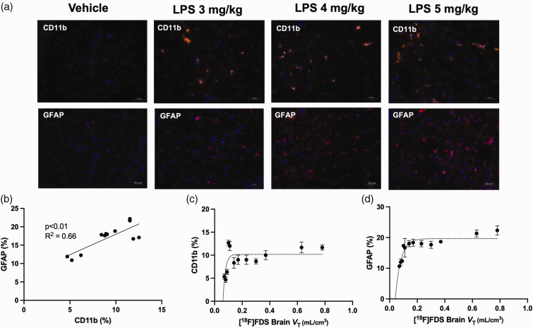
Figure 4.
Expression of glial cell markers relative to blood-brain-barrier (BBB) permeability, assessed using [18F]2-fluoro-2-deoxy-sorbitol ([18F]FDS) PET imaging in mice treated with increasing doses of lipopolysaccharide (LPS). The percentage of GFAP and CD11b-positive cells was measured using immunohistochemistry in the brain of 12 mice that received different doses of lipopolysaccharide saline (vehicle, n = 2) or LPS 3 mg/kg (n = 3), LPS 4 mg/kg (n = 4), or LPS 5 mg/kg (n = 3). Representative immunohistochemistry slices obtained in each condition are shown in a. Correlation between expression of CD11b and GFAP, expressed as a percentage of total nuclei from resected brain slices as markers of microglia or astrocytes, respectively, are shown in b. Correlation between the total brain distribution of [18F]FDS (VT), used for quantitative estimation of BBB permeability and corresponding expression CD11b and GFAP obtained in the same individuals are shown in c and d, respectively.

