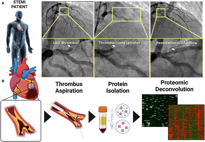
Figure 1. Coronary thrombus aspiration during STEMI provides the serological resource for high‐throughput proteomic assessment.

A, Angiographic depiction of an occlusive thrombus in the left anterior descending artery (left panel) and subsequent insertion of thrombectomy catheter with clot aspiration/restoration of blood flow (middle and right panels). B, Clots extracted via a coronary aspiration catheter (left 3 panels) were subjected to high‐throughput proteomic assessment to uncover the protein content of coronary blood at the time of reperfusion in STEMI. LAD indicates left anterior descending artery; and STEMI, ST‐segment–elevation myocardial infarction.
