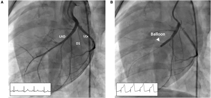
Figure 2. Angiography of the porcine heart MI model.

A, Preinfarction angiogram. Distribution of coronary artery blood flow along the anterior portion of the left ventricle is determined for proper balloon placement in the mid‐LAD. B, The angioplasty balloon is inflated to completely occlude the coronary artery as documented by repeat angiography and ST deviation on ECG monitoring. D1 indicates first diagonal branch; LAD, left anterior descending artery; and LCX, left circumflex artery.
