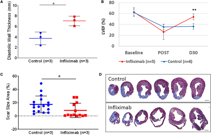
Figure 10. Late‐phase porcine myocardial infarction.

A, Apical anteroseptal wall thickness was obtained from intracardiac echocardiography. Evaluation revealed an increase in anterior wall thickness in diastole (7.07±0.91 mm for postinfarction infliximab treated [n=3] and 3.73±1.22 mm for control [n=3]; P<0.05) 4 weeks after MI. B, LVEF was obtained from intracardiac echocardiography at baseline (62.7±8.1 vs 62.0±6.5%) and immediately after MI (26.0±13.7 vs 35.3±5.3%), in infliximab‐treated (n=5) and control (n=6) cohorts. Preservation of LVEF at 30 days after MI was observed in the infliximab cohort (53.9±5.4 vs 36.2±5.3%; P<0.001). C, Systemic infliximab treatment (n=3) resulted in reduced scar size and diminished hypertrophic adverse remodeling in the left ventricle compared with untreated infarcted hearts (n=3) 4 weeks after MI (8.31±10.9% for infliximab vs 17.41±12.5% for control; P<0.05). Data points depict scar size area measurements from histology sections. D, Representative histological cross‐sections of pig hearts 4 weeks after infarction stained with Masson's trichrome to visualize collagen (blue) and myocardium (red/purple). Analysis performed with nonparametric statistical Kruskal–Wallis test. Asterisks indicate statistical significance. Scale bar represents 10 mm. LVEF indicates left ventricular ejection fraction; and MI, myocardial infarction.
