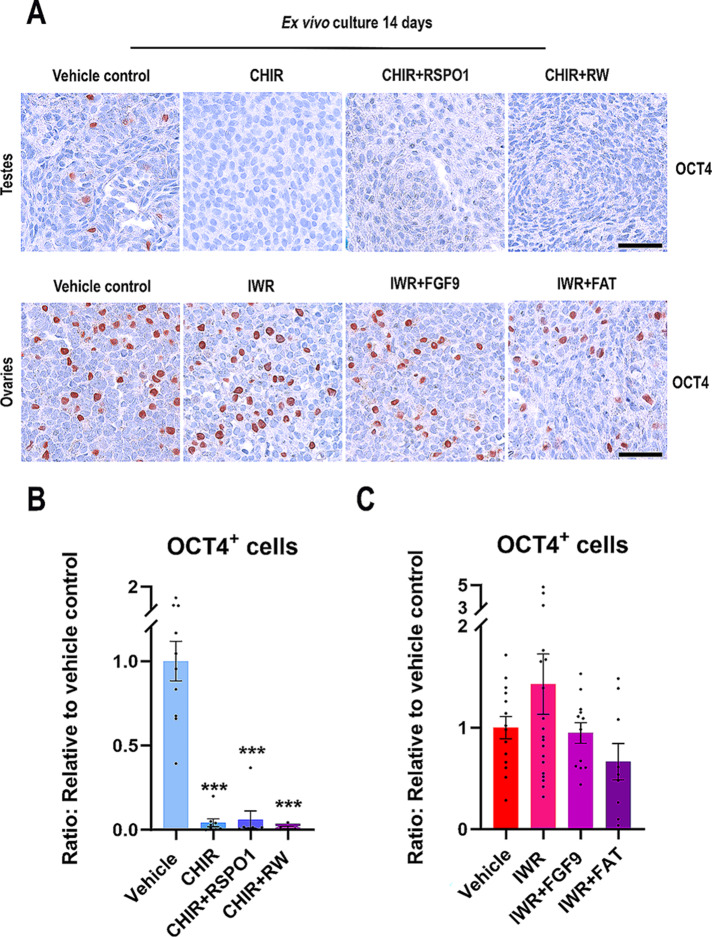
Fig. 8.
Manipulation of WNT/β-catenin signalling in ex vivo cultures of human fetal gonads severely reduce the number of germ cells in testes but not ovaries. (A) Representative images of OCT4 immunostaining in ex vivo cultured fetal testes treated with CHIR (3 µM) or CHIR + RW: CHIR (3 µM) + RSPO1 (100 ng/ml) + WNT4 (100 ng/ml) and ex vivo cultured fetal ovaries treated with IWR (1µM), IWR + FGF9: IWR-1 (1µM) + FGF9 (50 ng/ml) or IWR + FAT: IWR-1 (1µM) + FGF9 (50 ng/ml) + Activin A (25 ng/ml) + Activin B (25 ng/ml) + TGFβ (25 ng/ml). Counterstaining was performed with Mayer’s haematoxylin; scale bar 50 μm. Age of fetal samples shown (at start of experiment): Vehicle control 9 + 0 PCW; CHIR 8 + 2 PCW; CHIR + RSPO1 7 + 2 PCW; CHIR + RW 9 + 0 PCW for testes samples and vehicle control 7 + 0 PCW; IWR 7 + 2 PCW; IWR + FGF9 7 + 1 PCW; IWR + FAT 7 + 6 PCW for ovarian samples. (B) Quantification of the number of OCT4+ cells/mm2 in ex vivo cultured fetal testes treated with CHIR (n = 8), CHIR + RSPO1 (n = 7) or CHIR + RW (n = 5). Results are shown as fold change compared to internal vehicle control with data presented as mean ± SEM with individual datapoints included. Asterisk indicates statistical significance compared to vehicle control with *** P < 0.001. (C) Quantification of the number of OCT4+ cells/mm2 in ex vivo cultured fetal ovaries treated with IWR (n = 19), IWR + FGF9 (n = 12) or IWR + FAT (n = 9). Results are shown as fold change compared to internal vehicle control with data presented as mean ± SEM with individual datapoints included

