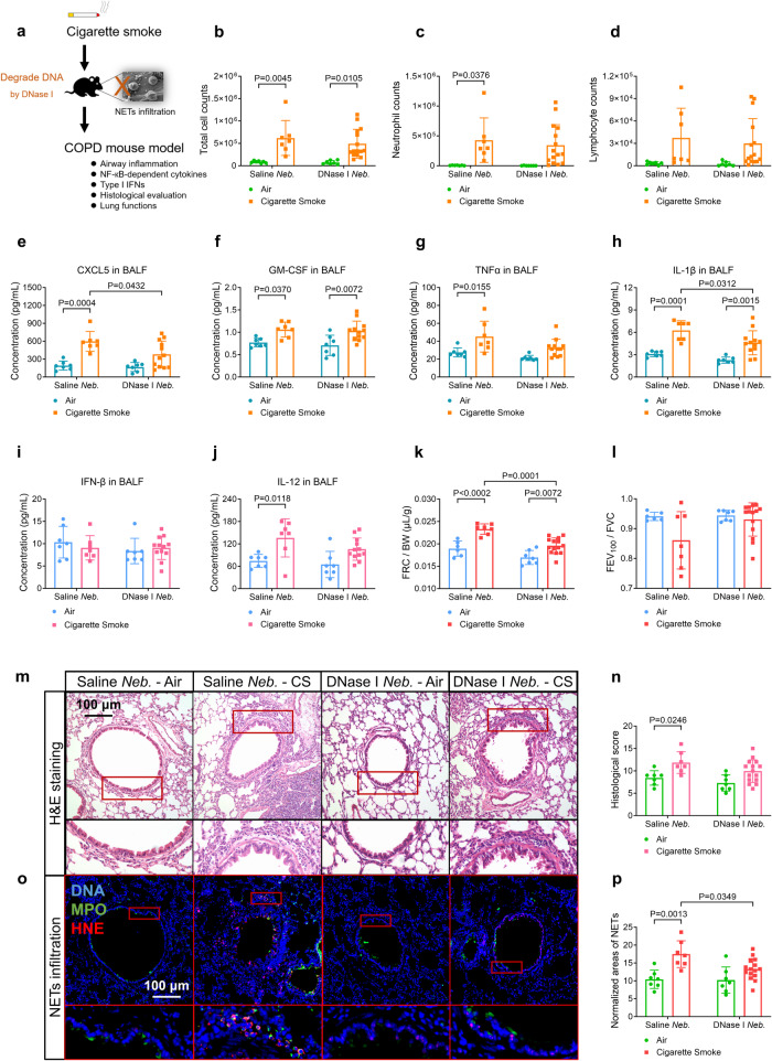
Fig. 7.
Cigarette smoke (CS)-exposed wild-type mice treated with the nebulization (Neb.) of deoxyribonuclease-I (DNase-I) reveal the decreased infiltration of neutrophil extracellular traps (NETs) and improved lung function, as compared with CS-treated saline Neb. mice. Statistical analysis: n = 7–15 for each bar in (b–l, n, p), data were presented as the mean ± standard deviation; Differences are assessed by the (b–l, n, p) two-way ANOVA analysis of variance, followed Tukey’s honest significant test; P < 0.05 represents a significant difference, the scattered samples and the p values are displayed in figures. a A brief outline for the experiments of the COPD mouse model (Supplementary Method 18, 19). b–d No significant changes of cell counts in bronchoalveolar lavage fluid (BALF) of CS-treated DNase-I Neb. mice are observed (Supplementary Method 22): b total cell counts, c neutrophil counts and d lymphocyte counts. e–h CS-treated DNase-I Neb. mice reveal the decreased levels of e C-X-C motif chemokine ligand 5 (CXCL5) and h interleukin 1β (IL-1β), but not that of f granulocyte-macrophage colony-stimulating factor (GM-CSF) or g TNFα (tumour necrosis factor-alpha) in the BALF (supplementary Method 22, 26). i, j No significant changes of type-I interferons (IFNs) level in the BALF of either CS-treated saline Neb. or DNase-I Neb. mice are observed: i IFN-β, j IL-12. k, l CS-treated DNase-I Neb. mice reveal alleviated emphysema, but not airflow limitation, in lung function tests (Supplementary Method 21) as evaluated by: k functional residual capacity/body weight (FRC/BW), l forced expiratory volume at 100 ms/forced vital capacity (FEV100/FVC). m Representative images of hematoxylin-eosin (H&E)-stained lung slices reveal that significant changes in the severity of airway inflammation are absent in the CS-treated DNase-I Neb. mice (scale bar: 100 μm, Supplementary Method 23), as summarised in n histological score. o Representative immunofluorescence images display the decreased infiltration of NETs (co-stained with DNA, MPO and Histone H3) in lung slices of the CS-treated DNase-I Neb. mice, compared with CS-treated saline Neb. mice (scale bar: 100 μm, Supplementary Method 24), as summarised in (p) normalised area of NETs

