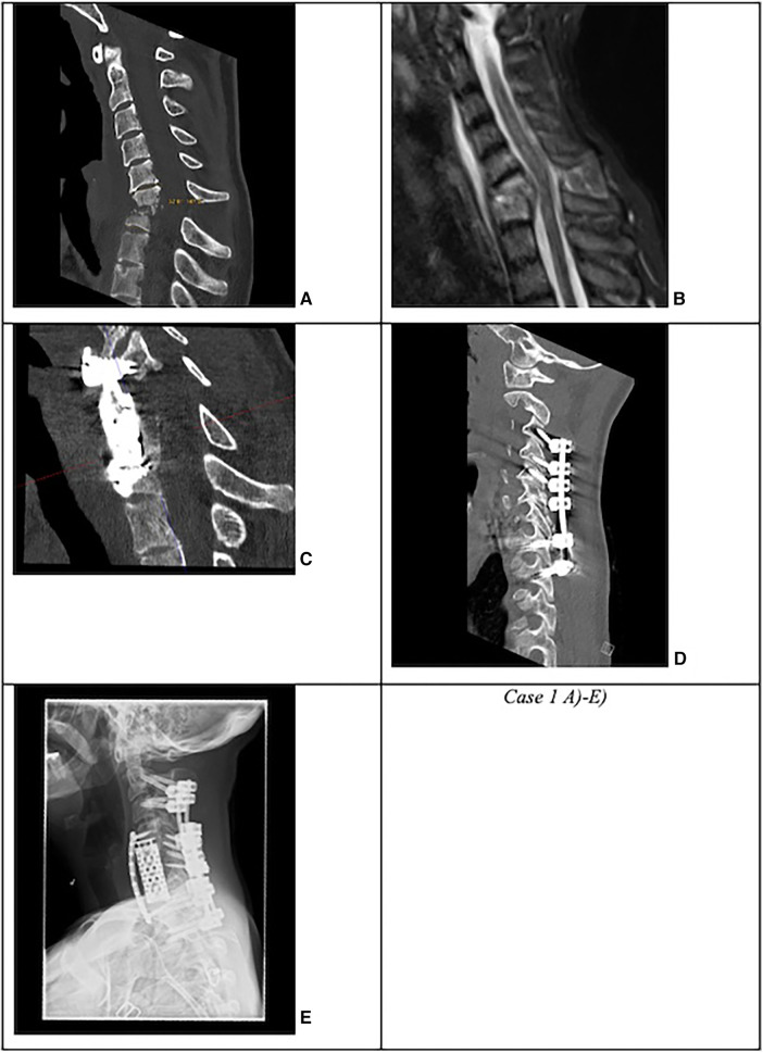
Figure 1.
Case 1: Complicated cervical spondylodiscitis in C6/7 with long-segment (three levels) posterior epidural abscess, kyphotic sagittal deformity with sagittal angle of 33°, and progressive osteomyelitis in a drug abuser. (A) CT and (B) MRI with C6/7 spondylodiscitis with 33° kyphotic deformity. (C) First revision surgery due to cage subsidence with dorsal spondylodesis extension to C4 and anterior plating. (D) Secondary lateral mass screw loosening in C3 and C4. (E) Second revision surgery with pedicle screw placement in C2 and C3.

