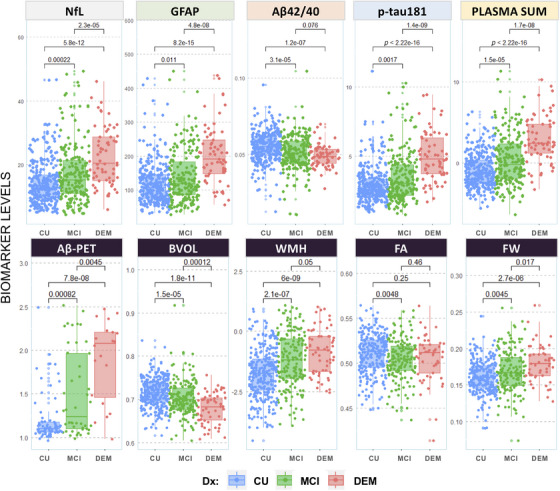
FIGURE 1.

Unadjusted group comparisons of plasma and neuroimaging measures by diagnostic status. Note that levels of plasma and neuroimaging biomarkers are compared across diagnostic groups (unadjusted; Dx: blue = CU; green = MCI; red = DEM). Aβ, amyloid beta; BVOL, brain volume (adjusted for intracranial volume); CU, cognitively unimpaired; DEM, dementia; DWI, diffusion weighted imaging; Dx, diagnosis; FA, fractional anisotropy; FW, freewater; GFAP, glial fibrillary acidic protein; MCI, mild cognitive impairment; NfL, neurofilament light; NODDI, neurite orientation dispersion and density imaging; PLASMA SUM = sum Z‐scored composite of NfL, GFAP, Aβ42/40, and p‐tau181; WMH, log‐transformed global white matter hyperintensity volume.
