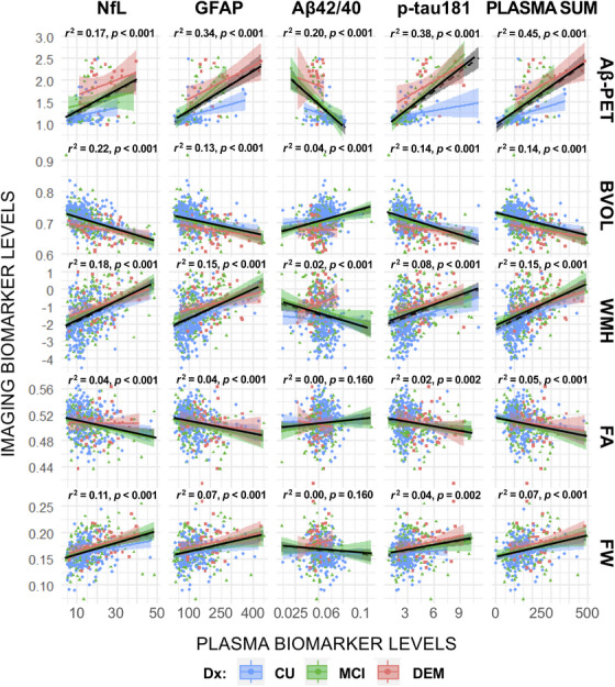
FIGURE 2.

Unadjusted linear relationships between plasma and neuroimaging biomarkers. Note that unadjusted bivariate associations between plasma and neuroimaging markers are presented. r2 statistics and p values represent lines of best fit (solid black line) for the full sample of participants (irrespective of consensus diagnosis). Plasma‐neuroimaging associations, stratified by clinical diagnosis, are provided for comparison (blue = CU; green = MCI; red = DEM). Plasma biomarkers are most strongly associated with Aβ‐PET deposition. Adj, linear models adjusted for comorbidities (eGFR, BMI); Aβ, amyloid beta; BVOL, brain volume (adjusted for intracranial volume); CU, cognitively unimpaired; DEM, dementia; base, baseline unadjusted models; DWI, diffusion weighted imaging; Dx, diagnosis; FA, fractional anisotropy; FW, free water; GFAP, glial fibrillary acidic protein; MCI, mild cognitive impairment; NfL, neurofilament light; NODDI, neurite orientation dispersion and density imaging; SUVr, standardized uptake volume ratio; WMH, log‐transformed global white matter hyperintensity volume; F‐Adj, adjusted models plus additional covariates (age, sex, race, education, APOE ε4 status, medications); F‐Adj*, fully adjusted models plus clinical diagnosis.
