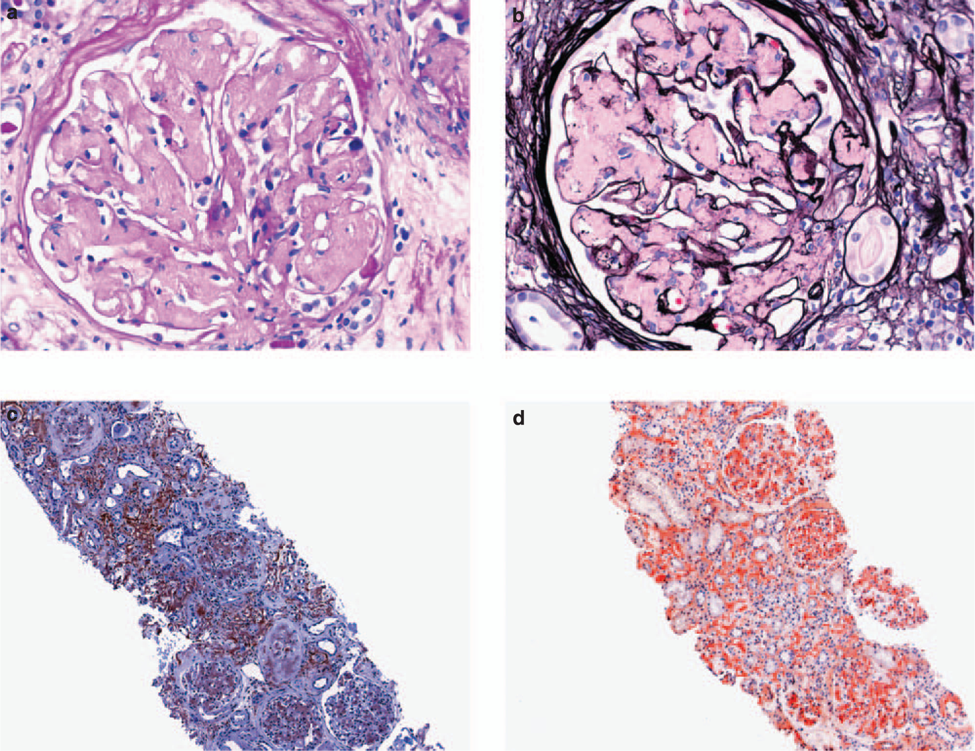
Figure 1 |. LECT2-associated renal amyloidosis (light microscopy).

(a) Extensive mesangial replacement by periodic acid-Schiff and (b) Jones methenamine silver-negative amorphous material (× 400). (c) Reactivity of glomerular and interstitial amyloid deposits with an anti-LECT2 antibody (immunoperoxidase technique, × 100). (d) Uptake of Congo red stain by interstitial and glomerular amyloid deposits (× 100). LECT2, leukocyte chemotactic factor 2.
