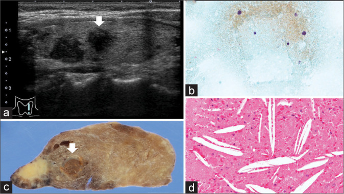
Figure 2:

Cystic follicular nodular disease. (a) Papillary thyroid carcinoma-suspected hypoechoic mass (white arrow) (Ultrasound B mode). (b) Cholesterol crystal and proteinaceous materials (Papanicolaou stain, x400). (c) Unilocular cystic lesion (white arrow). (d) Cholesterol crystal and proteinaceous materials (Hematoxylin and Eosin stain, x200).
