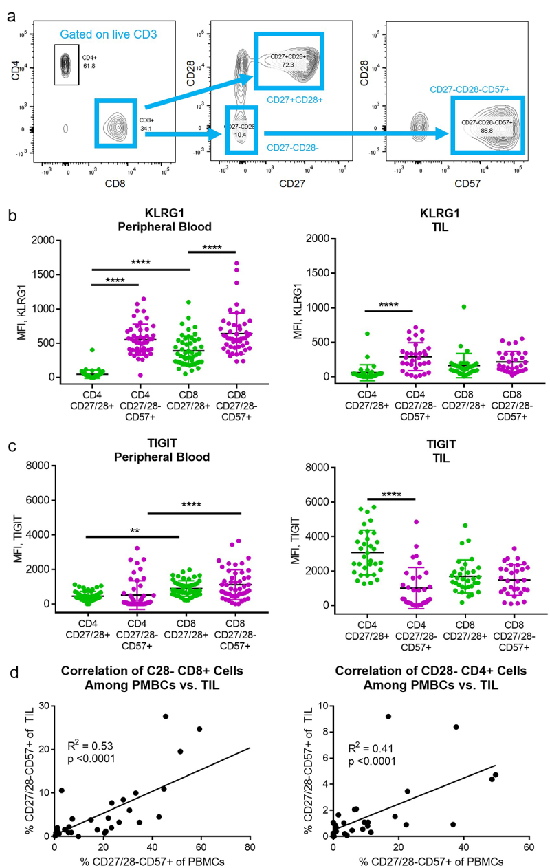
Figure 1.

CD27/28-CD57+ T cells and their CD27/28+ counterparts were isolated from the peripheral blood and tumor tissue of HNSCC patients. (a) Flow cytometry gating strategy used for CD8+ and CD4+ subsets. (b) KLRG1 expression among T cell subsets in the peripheral blood (left) and tumor infiltrating lymphocytes (TIL, right). MFI, mean fluorescence intensity. (c) TIGIT expression among T cell subsets in the peripheral blood (left) and TIL (right). (d) Correlation (Pearson correlation coefficient, squared) between percent of CD27/28-CD57+ T cells in the peripheral blood versus TIL for CD8+ subsets (left) and CD4+ subsets (right). Data in B and C represent mean ± one SD. **p < .01, ***p < .001, ****p < .0001 by two-way ANOVA with post hoc Tukey analyses.
