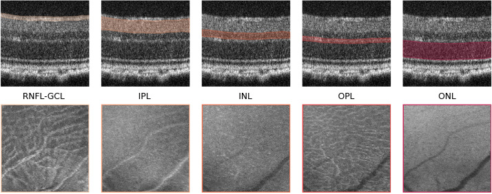
Figure 2.
Mean-value fundus (MVF) images. Example of MVF images (bottom) computed from the retinal nerve fiber layer and ganglion cell layer complex (RNFL-GCL), and the inner plexiform layer (IPL), inner nuclear layer (INL), outer plexiform layer (OPL), and outer nuclear layer (ONL). On top, the respective layers used for MVF computation are highlighted.

