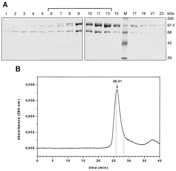
FIG. 2.
Analysis of RSV RT αD181NPol after elution from a heparin-Sepharose column. (A) SDS-PAGE of the eluted fractions. Proteins were detected by Coomassie staining. Lane M, protein molecular mass markers. The bracket indicates the pooled fractions. (B) HPLC size exclusion chromatography of the pooled fractions of αD181NPol (37). The peak with a retention time of 26.01 min corresponds to a molecular mass of ∼175 kDa. The retention times of homodimeric Pol (25.86 min) and α (28.26 min) were determined in separate runs with the corresponding enzymes and are indicated by dotted lines. The molecular mass of αD181NPol was determined using molecular mass standard proteins from U.S. Biochemical Corp. in the same buffer.

