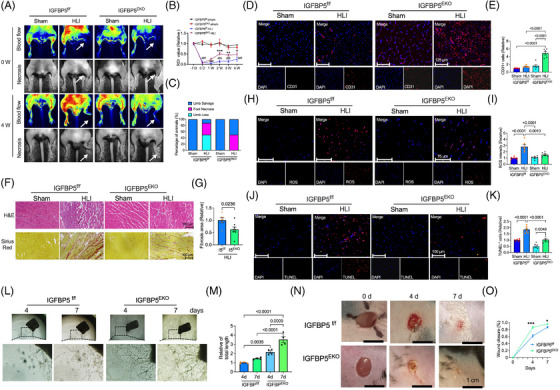
FIGURE 2.

Endothelial cell (EC)‐specific deletion of insulin‐like growth factor‐binding protein 5 (IGFBP5) attenuates the damage of the ischaemic hindlimb in mice. (A) Representative blood flow images of laser Doppler‐based tissue perfusion system in CDH5‐Cre‐IGFBP5flox/flox (IGFBP5EKO) and IGFBP5flox/flox (IGFBP5f/f) mice after ischaemia induction (0 W) and 4 weeks after ischaemia. (B) Quantification of blood flow in the hindlimb of IGFBP5EKO and IGFBP5f/f mice before (−1 D) and after ischaemia induction at 0, 1, 2, 3 and 4 weeks (n = 8 in each group). ### p < .001 compared to IGFBP5f/f‐sham; * p < .05, ** p < .01 and *** p < .001 compared to IGFBP5f/f‐HLI. (C) Percentage of limb salvage, foot necrosis and limb loss in IGFBP5EKO and IGFBP5f/f mice treated with sham or hindlimb ischaemia (HLI) (n = 8 in each group). (D) Representative immunofluorescence staining images and (E) quantification of CD31 (red) in gastrocnemius of hindlimb from IGFBP5EKO and IGFBP5f/f mice treated with sham or HLI (n = 5 in each group). (F) Representative haematoxylin‒eosin and picrosirius red staining images and (G) quantification of fibrosis area in gastrocnemius of hindlimb from IGFBP5EKO and IGFBP5f/f mice treated with sham or HLI (n = 5 in each group). (H) Representative immunofluorescence staining images and (I) quantification of dihydroethidium (DHE) staining for reactive oxygen species (ROS) in gastrocnemius of hindlimb from IGFBP5EKO and IGFBP5f/f mice treated with sham or HLI (n = 5 in each group). (J) Representative immunofluorescence staining images and (K) quantification of TUNEL staining in gastrocnemius of hindlimb from IGFBP5EKO and IGFBP5f/f mice treated with sham or HLI (n = 5 in each group). (L) Representative microscopy images of aortic sprouting assay and (M) quantification of tube length of the aorta from IGFBP5EKO and IGFBP5f/f mice at 4 and 7 days after being placed in plates with Matrigel. (N) Representative images and (O) quantification of cutaneous wound healing in IGFBP5EKO and IGFBP5f/f mice at 0, 4 and 7 days after wound induction (* p < .05, *** p < .001 vs. IGFBP5f/f).
