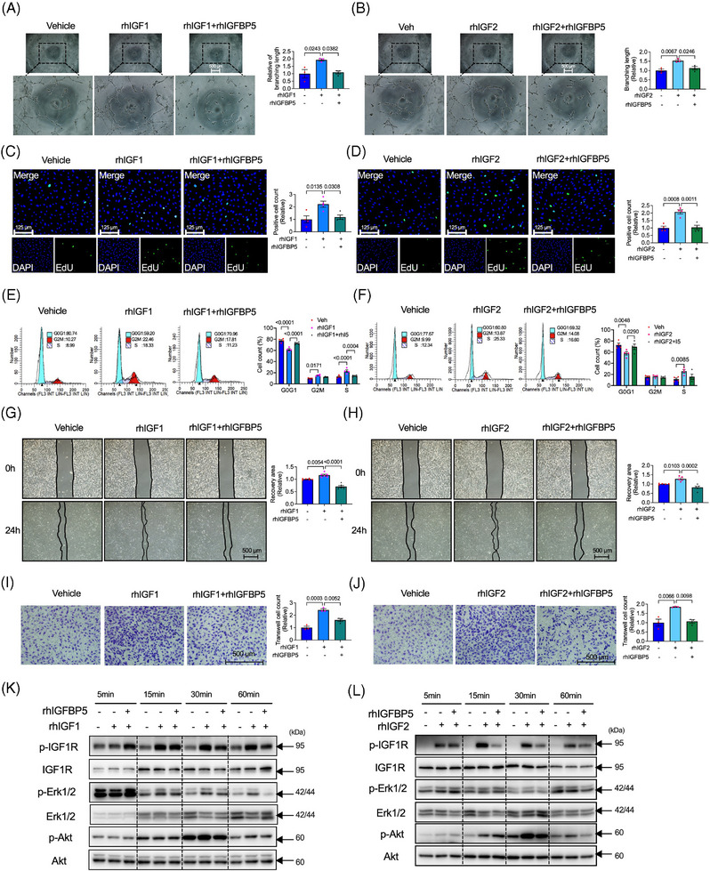
FIGURE 5.

Recombinant human insulin‐like growth factor‐binding protein 5 (IGFBP5) restrains IGF1‐ or IGF2‐induced tube formation, cell proliferation and migration. (A) Representative tube formation images and quantification of human umbilical vein endothelial cells (HUVECs) treated with rhIGF1 in the presence or absence of recombinant human IGFBP5 (rhIGFBP5) (n = 5 in each group). (B) Representative tube formation images and quantification of HUVECs treated with rhIGF2 in the presence or absence of rhIGFBP5 (n = 5 in each group). (C) Representative immunofluorescence images and quantification of 5‐ethynyl‐2′‐deoxyuridine (EdU, green)‐stained HUVECs treated with rhIGF1 in the presence or absence of rhIGFBP5 (n = 5 in each group). (D) Representative immunofluorescence images and quantification of EdU (green)‐stained HUVECs treated with rhIGF2 in the presence or absence of rhIGFBP5 (n = 5 in each group). (E) Representative images and quantification of flow cytometry for cell cycle of HUVECs treated with rhIGF1 in the presence or absence of rhIGFBP5 (n = 5 in each group). (F) Representative images and quantification of flow cytometry for cell cycle of HUVECs treated with rhIGF2 in the presence or absence of rhGFBP5 (n = 5 in each group). (G) Representative images and quantification of wound healing assay of HUVECs treated with rhIGF1 in the presence or absence of rhIGFBP5 (n = 5 in each group). (H) Representative images and quantification of wound healing assay of HUVECs treated with rhIGF2 in the presence or absence of rhIGFBP5 (n = 5 in each group). (I) Representative images and quantification of transwell assay of HUVECs treated with rhIGF1 in the presence or absence of rhIGFBP5 (n = 5 in each group). (J) Representative images and quantification of transwell assay of HUVECs treated with rhIGF2 in the presence or absence of hIGFBP5 (n = 5 in each group). (K) Representative images of Western blotting assay‐detected time‐course of p‐IGF1R, IGF1R, p‐Erk1/2, Erk1/2, p‐Ak and Akt expression of HUVECs treated with rhIGF1 in the presence or absence of rhIGFBP5. (L) Representative images of Western blotting assay‐detected time‐course of p‐IGF1R, IGF1R, p‐Erk1/2, Erk1/2, p‐Ak and Akt expression of HUVECs treated with rhIGF2 in the presence or absence of rhIGFBP5.
