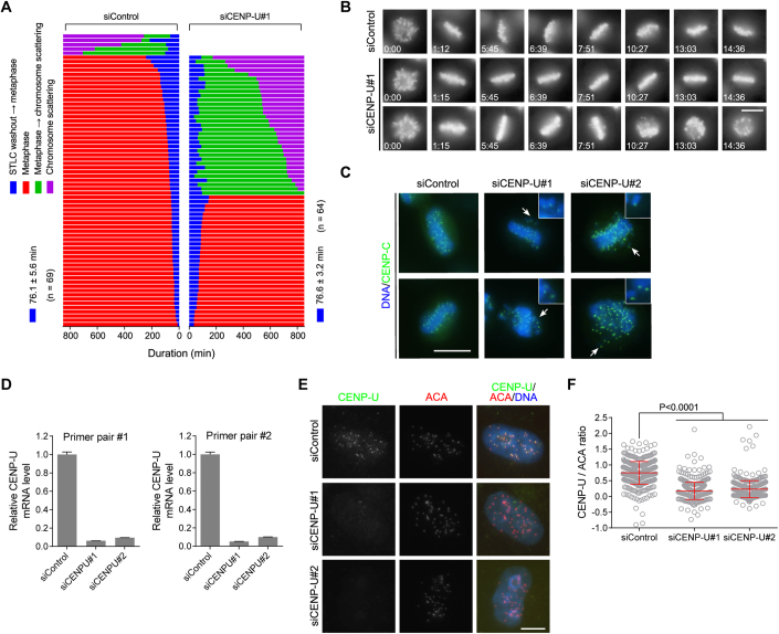
Figure EV1. CENP-U strengthens centromeric cohesion and promotes metaphase sister-chromatid cohesion.
(A, B) HeLa cells stably expressing H2B-GFP were transfected with control siRNA or CENP-U siRNA, followed by synchronization in S-phase with thymidine treatment for 20 h, and then released into fresh medium. At 7 h after thymidine release, cells were treated for 5 h with STLC, then mitotic cells were collected and released into fresh medium containing MG132 followed by live imaging of mitosis progression for 879 min. The time from STLC washout to metaphase chromosome alignment, and from metaphase to chromosome scattering, was determined and profiled (A). The selected frames of the movies are shown (B). The time stated in hours: minutes. See Movies EV1, EV2. (C) HeLa cells were transfected with control siRNA or CENP-U siRNA. At 48 h after siRNA transfection, cells were treated with MG132 for 6 h and then stained with the CENP-C antibody and DAPI. Example images are shown. Arrows point to misaligned chromosomes with single CENP-C foci. (D) HeLa cells were transfected with control siRNA or CENP-U siRNA. At 48 h post-transfection, total RNA was extracted and subjected to quantitative RT-PCR analysis using two pairs of CENP-U primers. The level of CENP-U mRNA in CENP-U-depleted cells relative to that in control HeLa cells were determined in three independent experiments. (E, F) HeLa cells were transfected with control siRNA or CENP-U siRNA. At 48 h post-transfection, cells were stained with anti-human centromere autoantibody (ACA) and the CENP-U antibody. Example images are shown (E). The immunofluorescence intensity ratio of CENP-U/ACA was determined from ~400 centromere regions in 20 cells, with statistics being performed using unpaired Student’s t-test (F). Data information: Means and SDs are shown (D, F). Scale bars, 10 µm (C, E).

