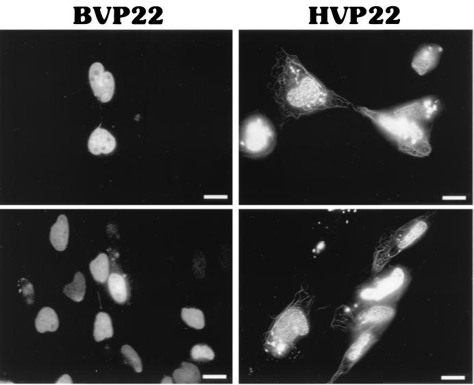
FIG. 6.
Distinctions in the nuclear localization patterns of BVP22 and HVP22. Fluorescence microscopy of transiently transfected D17 cells revealed a marbled pattern of nuclear staining by BVP22 and a speckled nuclear staining by HVP22. Frequently, BVP22-stained nuclei would be attached by filaments. Scale bar, 2 μm.

