
FIGURE 6.
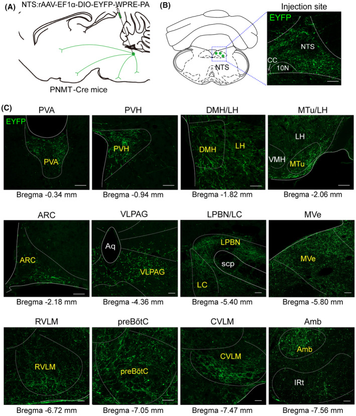
Projections of NTSPNMT neurons. (A) Schematic of anterograde tracing strategy of by injection of a recombinant anterograde vector to the NTS of PNMT‐Cre mice. (B) Typical images showing the expression of fluorescent PNMT neurons (green) in the NTS. Scale Bar, 100 μm. (C) Example distribution of axon terminals of NTSPNMT neurons. Scale bars, 100 μm. Amb, ambiguus nucleus; Aq, aqueduct (Sylvius); ARC, arcuate nucleus; CVLM, caudal ventrolateral medulla; DMH, dorsomedial hypothalamus; IRt, intermediate reticular nucleus; LC, locus coeruleus; LH, lateral hypothalamic area; LPBN, lateral parabrachial nucleus; MTu, medial tuberal nucleus; MVe, medial vestibular nucleus; preBötC, pre‐Bötzinger complex; PVA, paraventricular thalamic nucleus, anterior part; PVH, paraventricular hypothalamus; RVLM, rostral ventrolateral medulla; scp, superior cerebellar peduncle; VLPAG, ventrolateral periaqueductal gray; VMH, ventromedial hypothalamic nucleus.