
FIGURE 7.
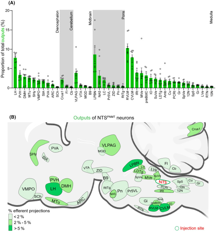
Summary of outputs from NTSPNMT neurons. (A) Statistical analysis of proportion of outputs distributed in each brain region to the total number of neurons in the whole brain. Brain areas are grouped into five general structures: the diencephalon, cerebellum, midbrain, pons, and medulla oblongata. Each error bar represents the SEM. Circles represent individual animal values (n = 8). (B) Summary of the major efferent outputs to NTSPNMT neurons. The depth of color represents the location and number of projections of NTSPNMT neurons. 10N, dorsal motor nucleus of vagus; 12N, hypoglossal nucleus; Amb, ambiguus nucleus; ARC, arcuate nucleus; B9, serotonin cells; Cb, cerebellum; Crus1, crus 1 of the ansiform lobule; Cu, cuneate nucleus; CVLM, caudal ventrolateral medulla; DMH, dorsomedial hypothalamus; Fl, flocculus; Gi, gigantocellular reticular nucleus; Gr, gracile nucleus; IO, inferior olive; IRt, intermediate reticular nucleus; LC, locus coeruleus; LDTg, laterodorsal tegmental nucleus; LH, lateral hypothalamic area; LPBN, lateral parabrachial nucleus; LVe, lateral vestibular nucleus; MGD, medial geniculate nucleus, dorsal part; MTu, medial tuberal nucleus; MVe, medial vestibular nucleus; NA5, noradrenalin cells; PCRt, parvocellular reticular nucleus; Pn, pontine reticular nucleus; Pr5VL, principal sensory trigeminal nucleus, ventrolateral part; preBötC, pre‐Bötzinger complex; PVA, paraventricular thalamic nucleus, anterior part; PVH, paraventricular hypothalamus; RTN, retrotrapezoid nucleus; RtTg, reticulotegmental nucleus of the pons; RVLM, rostral ventrolateral medulla; SCh, suprachiasmatic nucleus; SHy, septohypothalamic nucleus; Sp5, spinal trigeminal nucleus, caudal part; SpVe, spinal vestibular nucleus; StA, strial part of the preoptic area; SuVe, superior vestibular nucleus; VLPAG, ventrolateral periaqueductal gray; VMPO, ventromedial preoptic nucleus; VTA, ventral tegmental area; ZID, zona incerta, dorsal part.