
FIGURE 9.
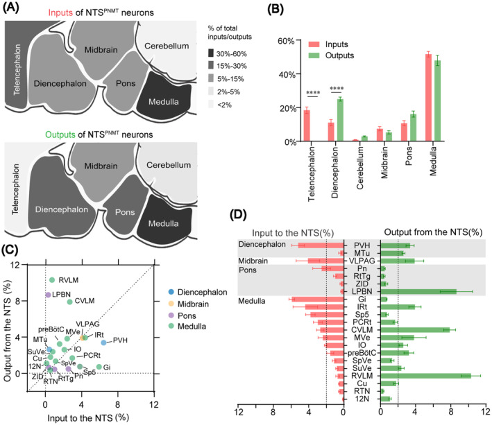
Comparison between inputs and outputs of NTSPNMT neurons. (A) Whole‐brain and output distributions of NTSPNMT neurons compared in six major structural general subdivision. Up: proportion of inputs to NTSPNMT neurons. Down: proportion of outputs from NTSPNMT neurons. (B) Percentage of total inputs to and outputs from the whole‐brain to NTSPNMT neurons. The data represent the mean ± SEM, with n = 4 for inputs and n = 8 for outputs. Levene's test followed by independent samples Student's ttests and Mann–Whitney U test (input vs. output: Telencephalon, p = 0.00001; Diencephalon, p = 0.00001; Cerebellum, p = 0.9859; Midbrain, p = 0.9544; pons, p = 0.1923; Medulla, p = 0.6140, ****p < 0.0001) were used to indicate statistical differences between inputs and outputs of NTSPNMT neurons in the same subdivision. (C) and (D) The percentage of inputs versus outputs in each region of typical nuclei that connected with the NTSPNMT neurons. Brain areas are grouped into four general structures: the diencephalon, midbrain, pons, and medulla. Error bar represents the SEM, with n = 4 for inputs and n = 8 for outputs. 12N, hypoglossal nucleus; Cu, cuneate nucleus; CVLM, caudal ventrolateral medulla; Gi, gigantocellular reticular nucleus; IO, inferior olive; IRt, intermediate reticular nucleus; LPBN, lateral parabrachial nucleus; MTu, medial tuberal nucleus; MVe, medial vestibular nucleus; PCRt, parvocellular reticular nucleus; Pn, pontine reticular nucleus; preBötC, pre‐Bötzinger complex; PVH, paraventricular hypothalamus; RTN, retrotrapezoid nucleus; RtTg, reticulotegmental nucleus of the pons; RVLM, rostral ventrolateral medulla; Sp5, spinal trigeminal nucleus, caudal part; SpVe, spinal vestibular nucleus; SuVe, superior vestibular nucleus; VLPAG, ventrolateral periaqueductal gray; ZID, zona incerta, dorsal part.