Fig. 1.
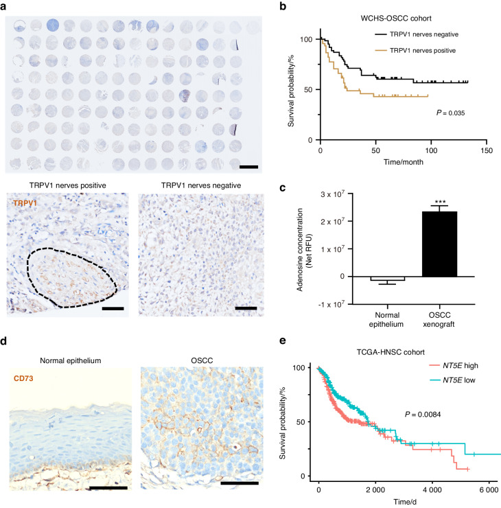
Presence of nociceptive nerves in adenosine-concentrated OSCC. a The upper panel presents the broad view of immunostaining of TRPV1 in the patient cohort collected by West China Hospital of Stomatology (WCHS). Scale bar: 2 mm. The lower panel denotes representative samples with and without TRPV1 neuronal infiltration (‘TRPV1 positive’ and ‘TRPV1 negative’). Scale bar: 50 µm. b Kaplan–Meier plot delineating survival probability for WCHS-OSCC patients stratified against TRPV1 neuron infiltration in their tumors, n = 111 patients. c Comparison of adenosine concentration in normal epithelium and HSC3 xenograft of mice, represented as net relative fluorescence unit (RFU). n = 3 mice. d Immunostaining of CD73 in human normal epithelium and human OSCC cells. Scale bars, 50 μm. e Kaplan–Meier plot delineating survival probability for TCGA-HNSC patients stratified against NT5E expression in their tumors. NT5E-high and NT5E-low groups are defined as above or below the median of NT5E expression. The TCGA-HNSC cohort database analyzed was updated to March 29th, 2023, n = 518 patients. Statistical analysis was conducted using unpaired Student’s t-test (c) and Log-rank test (b, e)

