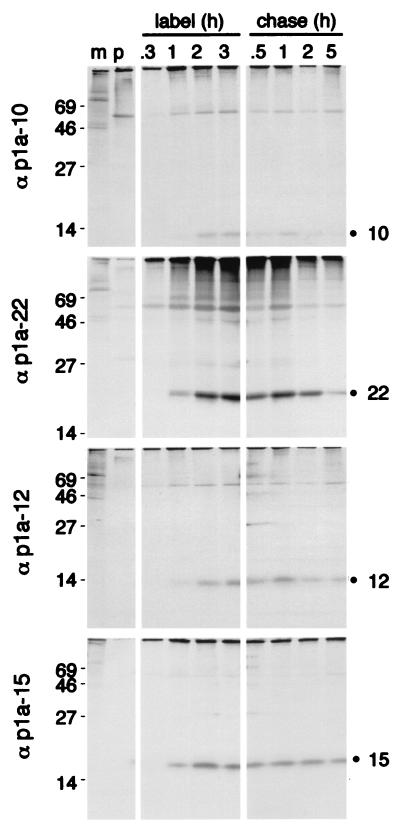
FIG. 3.
Pulse-label and pulse-chase translation of p1a-10, -22, -12, and -15. For pulse-label translation (label), DBT cells were mock infected (m) or infected with MHV-A59 for 5.5 h and were incubated with [35S]Cys-Met for 0.3, 1, 2, or 3 h prior to immunoprecipitation with the antibodies as indicated to the right of each gel. p denotes immunoprecipitation with preimmune serum. For pulse-chase translation (chase), DBT cells were radiolabeled beginning at 5.5 h p.i. with [35S]Cys-Met for 90 min prior to addition of excess cold methionine and cycloheximide for the times indicated above each well. Following harvesting and immunoprecipitation of the cells, label and chase samples for each antibody were run on the same SDS–10 to 20% gradient polyacrylamide gel, followed by fluorography. Molecular mass markers (kilodaltons) are shown to the left of the gels, and the locations of the p1a-10, -22, -12, and -15 proteins are indicated to the right of the gels.

