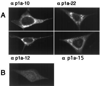
FIG. 4.
Localization of p1a-10, -22, -12, and -15 in MHV-infected cells. MHV-A59-infected or mock-infected DBT cells were fixed and prepared for immunofluorescence microscopy as described in Materials and Methods, using the antibodies as noted by each frame. Cells were imaged on a Zeiss LSM 410 confocal microscope using a 488-nm laser with acquisition in the LSM software. The images are single confocal slices using a 63× objective. Image processing (brightness and contrast) was performed in Photoshop 5.0. A representative mock-infected cell probed with αp1a-22 is shown in panel B. αp1a-10, -12, and -15 resulted in a similar lack of background staining in the mock-infected cells.

