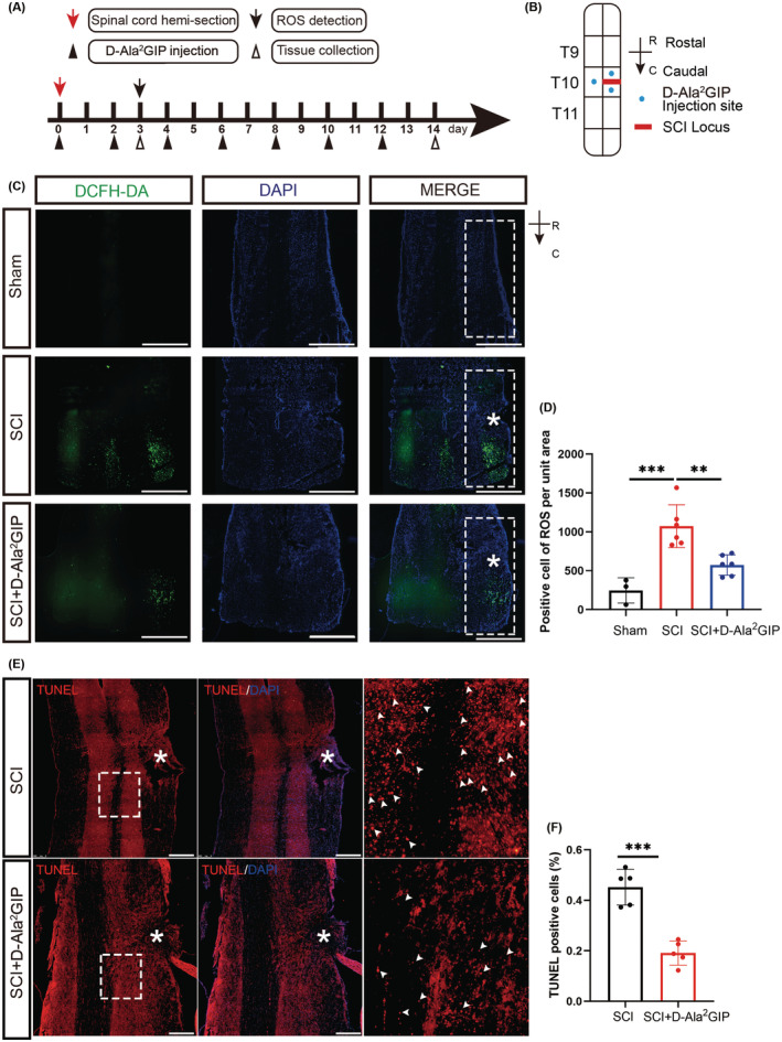
FIGURE 3.

Effect of GIP on oxidative stress in rats with spinal cord hemisection injury. (A) Timeline of spinal cord hemisection injury, D‐Ala2GIP administration, and tissue collection. (B) Model of GIP injection in spinal cord injury. Spinal cord hemisection was conducted at T10. For saline and D‐Ala2GIP treatments, each rat was injected with 3 μL saline or D‐Ala2GIP at three sites. R: Rostral, C: Caudal. (C, D) D‐Ala2GIP decreased ROS level after spinal cord hemisection injury for 3 days. The white “*” shows the injury sites and the white box shows the area for statistics of ROS signal. Scale Bar = 500 μm. Statistical results of ROS‐positive cells were shown. n = 3 for sham group, n = 6 for SCI group and SCI + GIP group. The data were shown as mean ± SE, and were analyzed by one‐way ANOVA, **p < 0.01, ***p < 0.001. (E, F) D‐Ala2GIP decreased the number of apoptotic cells after spinal cord hemisection injury for 14 days. The white “*” shows the injury sites, the white arrows are representative TNUEL‐positive signals, and the images on the right are magnified views of the boxed area. Scale Bar = 250 μm, Statistical result of TUNEL mean fluorescence intensity after spinal cord hemisection injury for 14 days. n = 5 for each group. The data were shown as mean ± SE, and were analyzed by Student's t‐test, ***p < 0.001.
