Fig. 1.

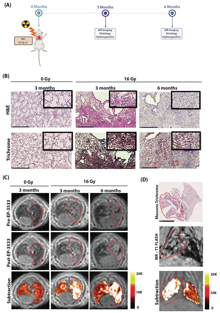
(A) Murine model of progressive radiation-induced lung injury. Mice underwent a single dose of 16 Gy to the right hemithorax and were imaged at either 3 or 6 months after irradiation. (B) Representative hematoxylin and eosin (H&E; top) and trichrome (bottom) stains of the right lung show normal lung architecture in unirradiated (0 Gy) mice. Mice at 3 months developed cellular infiltrate, alveolar thickening, consolidation, and collagen deposition. By 6 months, significant fibrosis and alveolar consolidation was seen. (C) Representative axial magnetic resonance (MR) images are shown before and after EP-3533 injection. Lungs of unirradiated mice (0 Gy) show low signal on ultrashort echo time MR. Irradiated mice show increasing right lung signal from 3 to 6 months due to consolidation, and these areas are enhanced after EP-3533 administration. False color subtraction images (40 minutes after EP-3533 injection — preinjection) overlaid on preinjection ultrashort echo time images demonstrate increased signal enhancement in irradiated lung. (D) Colocalization of lung injury in the irradiated right hemithorax on pathology and coronal MR images. Diffuse injury throughout the right lung but not in the left lung is seen on Masson’s trichrome and MR-T1FLASH images. The false color subtraction image illustrates substantial signal enhancement in the right lung but not in the nonirradiated left lung. Abbreviation: XRT = Radiation treatment.
