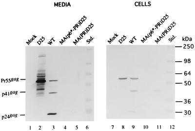
FIG. 2.
Expression and processing of the chimeric proteins. 293T cells were transfected with the designated constructs. At 48 h posttransfection, cells and supernatants were collected for protein analysis. Supernatant samples (lanes 1 to 5) corresponding to 50% of the total samples and cell samples (lanes 7 to 11) corresponding to 5% of the total samples were fractionated by sodium dodecyl sulfate–10% polyacrylamide gel electrophoresis and electroblotted onto a nitrocellulose filter. HIV p24gag and p24gag-associated chimeric proteins were detected with mouse anti-p24gag monoclonal antibody at a 1:5,000 dilution, followed by a secondary alkaline phosphatase-conjugated sheep anti-mouse antibody at a 1:5,000 dilution, and alkaline phosphatase activity was determined. Positions of standard (Std.) molecular size markers (lanes 6 and 12) are indicated on the right, and those of HIV Gag proteins Pr55, p41, and p24 are shown on the left.

