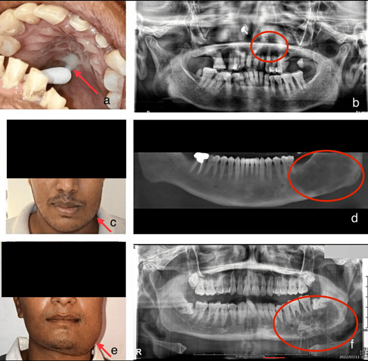
Figure 1. Figures showing clinical and radiographic images.
a) Purulent white discharge in the palatal region of fungal osteomyelitis - mucormycosis
b) Orthopantomogram revealing a hazy radiolucency extending from 21 to 25 region in the maxilla of fungal osteomyelitis - mucormycosis
c) Swelling involving the left lower side of the face - ossifying fibroma
d) Cone beam computed tomography (CBCT) revealing a mixed lesion with indistinct borders extending from the distal part of 36 and extending posteriorly to the ramus of the mandible - ossifying fibroma
e) Facial asymmetry involving the left lower part of the face - osteosarcoma
f) Orthopantomogram revealing a typical sunburst appearance of a mixed osteolytic lesion on the left posterior mandible with indistinct borders - osteosarcoma

