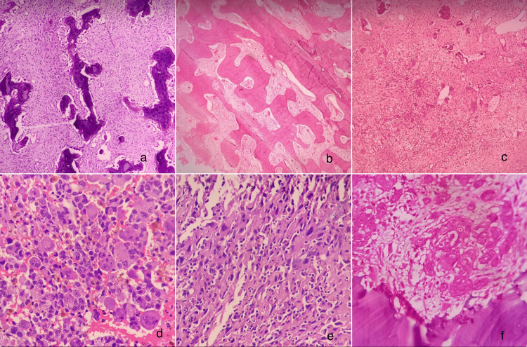
Figure 2. Photomicrographs.
a) Ossicles of bone within a cellular fibrous connective tissue stroma - ossifying fibroma (10x, H&E)
b) Bony trabeculae in Chinese letter pattern - fibrous dysplasia (10x, H&E)
c) Cellular connective tissue with bone elements and vascular channels - cherubism (4x, H&E)
d) Sheets of malignant cells with areas of hemorrhage - osteosarcoma (40x, H&E)
e) Diffuse proliferation of malignant cells - osteosarcoma (40x, H&E)
f) Islands of malignant epithelial cells invading the bone - squamous cell carcinoma invading the bone (10x, H&E)

