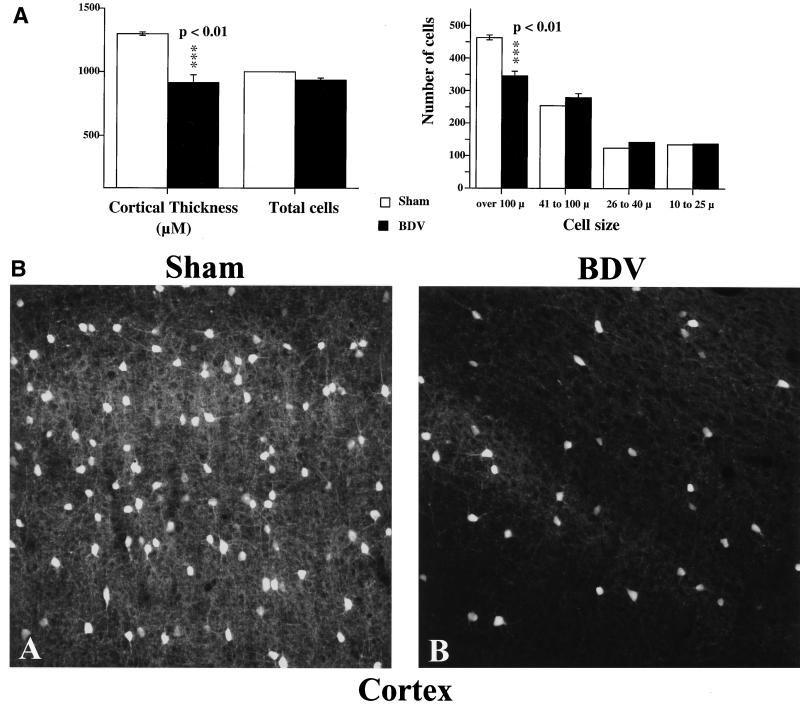
FIG. 5.
Cell loss in the cortex of neonatally BDV-infected animals, 45 days p.i. (A) Morphometric analysis. Results are expressed as mean ± SEM. Averages are based on results from five each sham-inoculated and rats with BDV PTI (two sections per animal). The level of statistical significance (determined by Mann-Whitney U test) is indicated by triple asterisk (P < 0.01). BDV-infected rats display significant cortical shrinkage (about 30%) and loss of cells with a diameter of >100 μm compared to noninfected age-matched controls. (B) Immunohistochemical staining for parvalbumin in the cortex of sham- and BDV-infected rats, 45 days p.i. Parvalbumin, a calcium-binding protein, labels GABA-ergic neurons in the cortex. Similar cortical areas are shown for each animal. Note the significant decrease in numbers of stained cells and processes in the infected animal. Magnification, ×250.

