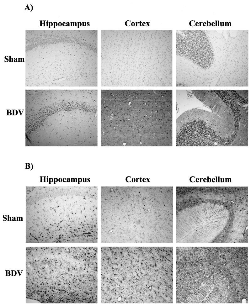
FIG. 6.
Detection of BDV antigen and GFAP expression in control and infected rat brains, 35 days p.i. (A) Expression of BDV nucleoprotein. Sections were immunostained with an antibody specific for BDV nucleoprotein. There is a strong nuclear staining in abundant pyramidal cells of the hippocampus and neocortex, as well as in Purkinje and granule cells of the cerebellum. Diffuse staining of the neuropil is also observed. There was no staining in sham-inoculated animals. Magnification, ×80. (B) Analysis of GFAP expression in similar fields. Neonatally infected rats display a significant increase in the number of GFAP-positive astroglial cells in the hippocampus (molecular layer and dentate gyrus), cortex, and cerebellum. Consistent with this activation, astrocytes are significantly hypertrophied in infected brains. Magnification, ×80.

