. 2024 Jun 16;15(1):2360196. doi: 10.1080/19491034.2024.2360196

Figure 1.

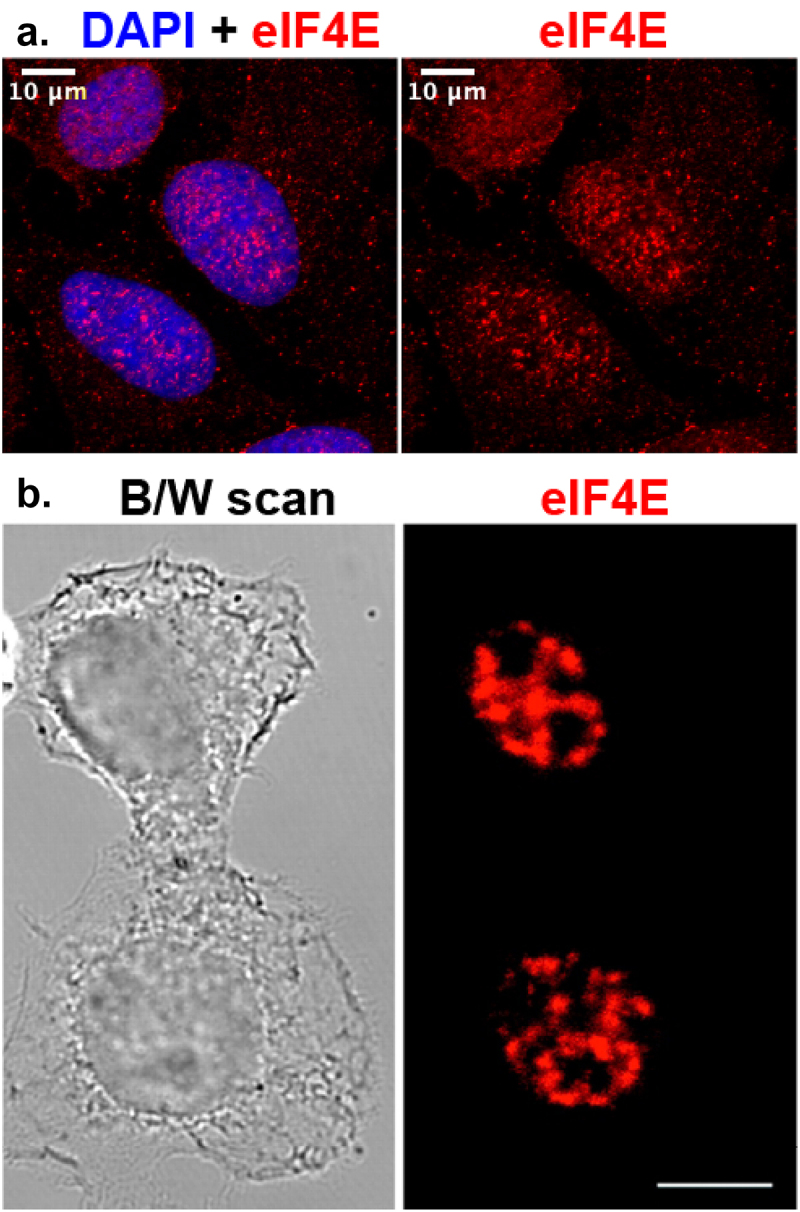
A. The top left panel shows DAPI staining of nuclei of three individual U2OS cells merged with the red signal of eIF4E staining, and on the right panel, the staining for eIF4E alone is seen in the same cells. Enrichment of eIF4E in the nuclei of cells is clear, cytoplasmic signal is also visible, showing a particulate and dispersed pattern through the nucleus and cytoplasm. B. The bottom left panel represents the phase contrast image of the two HeLa cells. The right panel represents eIF4E staining of the same cells showing predominantly nuclear localization.

A, Single section of confocal imaging of U2OS cells stained for eIF4E (sc-271480 anti-eIF4E antibody, red) and DAPI (blue, nuclear dye) showing nuclear and cytoplasmic localization. Left panel shows the overlap between DAPI and eIF4E staining. The right panel shows eIF4E signal alone. B, Confocal imaging of eIF4E in the nucleus of HeLa cells with the antibody 10C6 from [101]. Reprint with permission from Dostie J, Lejbkowicz F, Sonenberg N. Nuclear eukaryotic initiation factor 4E (eIF4E) colocalizes with splicing factors in speckles [99]. White bar = 10μm.